Basics of Visual System Anatomy
Introduction
Vision is the ability to use our eyes to understand the physical world without touching it, and to use visual information for thinking and for calculating muscle movements.
Vision relies upon one’s understanding of where the body is in space, how it is positioned, and where other objects are in relation to the viewer. Only once this is understood can we direct our eyes towards objects of interest. In human neurology, this all requires the integration of some 65% of the brain’s functions. A great part of the purpose of our brainstem functions, it seems, is to simply direct the head and eyes so we can ‘read’ the environment by using the shorthand of vision. Unlike early ‘somatic’ vision, where as infants we use the hands, lips, and tongue to understand objects, mature developed vision is much more potent. As adults, we use the highly evolved and broadened scope of vision to not only see the environment, but to read it, and anticipate it.
Most of us are familiar with the idea of an eye chart and how this can be used as a test of eyesight. This simple measure, visual acuity, provides useful information but is very limited in what it does tell us. Sight tests in schools, that is, distance visual acuity tests, ironically more often than not fail those children who are at a relative advantage in the classroom (nearsighted children), while passing most children who have significant visual impediments to learning. They are inefficient in assessing visual impediments given the complex nature of the visual process.
For developmental professionals, proper visual functional assessment and management is critical to diagnosing underlying conditions because many behavioural concerns are rooted in visual impediments alone, or have vision as a significant contributing factor. Also, addressing visual dysfunction opens the door to accelerated progress in collateral therapies by providing a stronger foundation for physical control, perception, and cognition.
This and the following two chapters provide a practical overview of some of the varied aspects of the visual system and function that can and will negatively impact upon behaviour when affected by developmental problems or pathology.
Basic Visual Anatomy
Vision is complex and integrated into some 65% of what our brains do. The eyes themselves are only one part of the visual process and are, obviously, the main tools used by vision to obtain sight-based information from the environment. Still, even if the eyes are closed, much of what the visual system does is still active; even if an individual becomes blinded, vision remains intact to a great degree because of its wide influence in the brain.
The visual system consists of internal structures, those we cannot see because they are inside the brain, and the external structures, namely the eyes. While the internal functioning of the visual system is important, we can really only infer what is happening by observing other behaviours, such as how a child responds to a test of visual memory, sequencing, or figure-ground, or by functional MRI, or by close study under a microscope. What is much simpler, more direct, and still very useful, is to study the details of the eyes’ themselves and how they move. What the eyes do is a direct function of what the brain is telling them to do, and likewise, the limits of the what the eyes are capable of doing will determine to a great degree what the brain will perceive. This discussion, then, will focus on the elements of the eyes and those muscles that help to move them and to focus images.
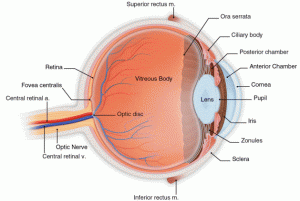
The eyes themselves are incredibly complex and a full description of their physiology and function is best left to books such as “Adler’s Physiology of the Eye” by Kaufman and Alm (Mosby, 2002), or “Clinical Anatomy and Physiology of the Visual System, 3rd Ed.”, by Remington (Elsevier, 2012). For now, a brief overview of some of the elements of the eye structure is worthwhile:
- Each eye’s movement is controlled by 6 muscles, the extraocular muscles.
- The eyes work independently of one another, but their movement is coordinated by different parts of the brain from brainstem to cortical areas.
- There are gross eye movements to quickly shift gaze or maintain gaze, and fine motor movements for very precise tracking of targets or moving quickly across text. Different movement types are controlled by different parts of the brain.
- Inside the eye, there are the intraocular muscles, those muscles primarily concerned with controlling the size of the pupil, and another set that controls focus.
- Each eye has an optic nerve that carries information to varied points in the brain, not just to the primary visual cortex.
- The retina, or the collection of light sensitive nerve fibers lining the inside of the eyes, is comprised of multiple layers of specialized cells.
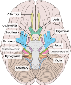
- Of the 12 cranial nerves (CN), 8 are somehow related to visual function, helping to integrate touch, sound, sight, balance, and muscle control. The optic nerve (CN II) is one of these (retinal input to brain centres), the others include: oculomotor (CN III, movement of the eyes, focusing / accommodation, pupil function), trochlear (CN IV, movement of the eyes), trigeminal (CN V, sensory feedback from the eyes and eye sockets), abducens (CN VI, eye movements), facial (CN VII, facial muscle control for squinting, marginal involvement in the visual process), vestibulocochlear aka auditory-vestibular (CN VIII, balance and auditory localization), and the accessory nerve (CN XI, movement of head/neck to orient).
Beyond the structure and function of the eyes themselves, there are numerous areas in most parts of the brain that are related to visual function and visual information processing. With 8 of 12 cranial nerves involved and dozens of interconnections inside the brain, it is easy to see that vision is very tightly integrated with most critical body functions. In a similar fashion, because the visual system is so tightly integrated to brainstem functions, for example, even mild trauma, such as whiplash or other mild traumatic brain injury (mTBI) can and will have important effects on visual function. (See Notes section on ‘cranial nerves’ above for more information.)
|
Schematic cross section of the human eye. |
The 12 Cranial Nerves as viewed on the inferior, or under, side of the brain. |
|
Extraocular muscles receive input from the brain to change the direction of the eyes and control fine eye movements.
|
There is much more to vision than anatomy, there are multiple neural and neuromuscular feedback loops that control our visual attention and perception, and how we use targeting to direct the eyes’ movement. To get a sense of how complex vision is, consider the following list of diagnostic procedures available and the domains they cover. This does not include more common psycho-educational tests that are in use in schools currently. Dr. Lea Hyvarinen’s, a prominent developmental ophthalmologist (and author the the LEA pediatric acuity symbols), has outlined a model for assessing the various visual functions as follows:
OCULAR MOTOR FUNCTIONS AND REFRACTION • Fixation, Saccades, Accommodation, Following • Strabismus, Nystagmus, Head and body control • Refractive errors, corrective spectacles and devises
VISUAL SENSORY FUNCTIONS • Visual acuity, near (single, line, crowded), distance, Grating acuity • Contrast sensitivity, optotype and grating tests • Colour vision, Visual adaptation to changes in luminance level, filter lenses • Figure in motion, Biological motion, Perception of motion at high speeds • Visual field: Extent, scotomas, Goldmann, flicker, automated campimetry
EARLY VISUAL PROCESSING • Perception of length and orientation of lines and objects • Figure-ground, object-background, stereovision, Vernier acuity • Short time visual memory, matching colours
IN THE VENTRAL STREAM/ INFEROTEMPORAL NETWORKS • Details in pictures, Noticing errors and missing details, Perception of textures and surface qualities • Recognition of familiar and unfamiliar faces, Facial expressions, Body language • Landmarks, Concrete objects, Pictures of concrete objects • Abstract pictures of objects of different categories, Abstract forms (letters, numbers) • Reading words and lines of texts, Optimal reading strategy • Comparison with pictures in memory, ‘Reading’ series of pictures • Visual problems in copying pictures from blackboard and/or at near • Crowding effect, Scanning lines of text
IN THE DORSAL STREAM/ PARIETAL NETWORKS •Awareness of surrounding space, directions and distances in space, Body awareness •Perception of near and far space, Orientation in space, map based, memorizing routes •Motion perception, Depth perception, Simultaneous perception •Eye-hand coordination, Grasping and throwing objects, Drawing, free hand •Copying from near/ from blackboard, motor planning and execution.
Introduction to LVT Quick Reference


